HEALTH HACK: Discover Types and Causes of Elephantiasis, and How to manage it.
Elephantiasis is a nematodes disease. Larva of microfilariae is the main source. Filarial worms are long and thin. It attacks the lymphatic system and subcutaneous connective tissue.
Filariasis has two main types named as
Onchocerca Volulus
Mansonella Streptocerca
These species of filaria inhabit in dermis and subcutaneous tissue. The cause of elephantiasis is Wuchereria bancrofti. This species occurs in Pakistan. Other species do not exist. Man is the definitive host while the Mosquito genus CULEX is the intermediate.
Filaria has separate sex. They live in pairs, therefore they block the lymphatic ducts resulting Elephantiasis.
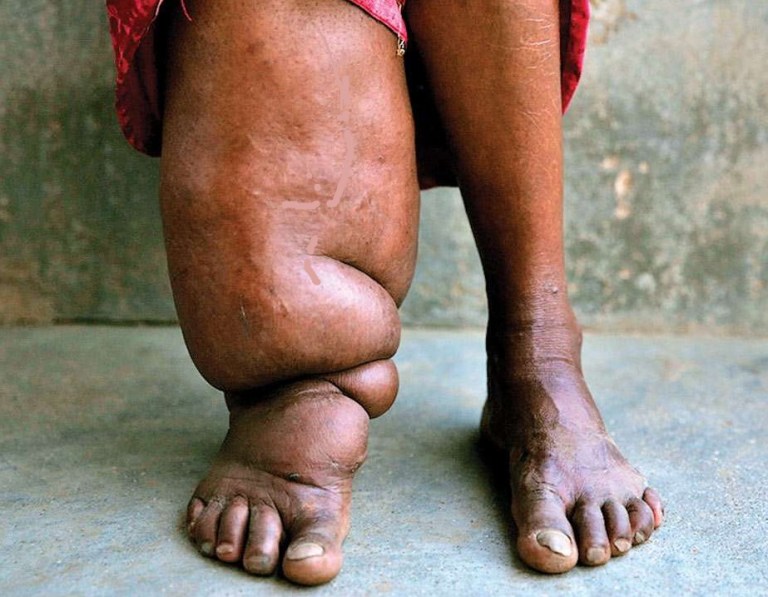
Elephantiasis symptoms
The life cycle of Filariasis
Culex mosquitoes take a blood meal from infected people. It ingest Microfilaria. This microfilaria penetrates into the midgut and goes to the thoracic muscle of the mosquito.
In the thoracic muscle this develops into the first stage, then third-stage larvae (Infective stage).
This L3 form larva migrates to the proboscis.
When this mosquito bites a healthy human, it transforms the L3 larvae. L3 forms inhabitants in lymphatic ducts and developing into adults.
Adults produce sheathed microfilaria into lymph and blood. It moves from the lymph duct to the bloodstream freely. Elephantiasis describe the causes and symptoms
Laboratory Diagnosis of Filariasis (Elephantiasis)
For the diagnosis of filarial infection, parasite demonstration is the accurate means. Blood should be collected around midnight (at this time parasites mostly present in the blood). There are three methods for parasite demonstration.
Thin Film Preparation
Thick Film Preparation
Concentration method
Thin Film Preparation method
Thin film smear prepared. Examine under microscope first at low power then at high power for more detail.
Thick Film Preparation
Prepare film and stain with Giemsa for better result. Hematoxylin and eosin stain can be parasite demonstrations. For this purpose, the smear should be air-dried and fixed with an al quantity of ether and alcohol for ten minutes. Dried and stained like histo specimen.
Concentration Method
In this method, blood should be centrifuged after collection in a tube containing 2% acetic acid. It should be mixed thoroughly. After centrifugation, the deposit examined under a coverslip. Here you will observe actively moving microfilaria.


